The integrated platform for organ-on-chip!
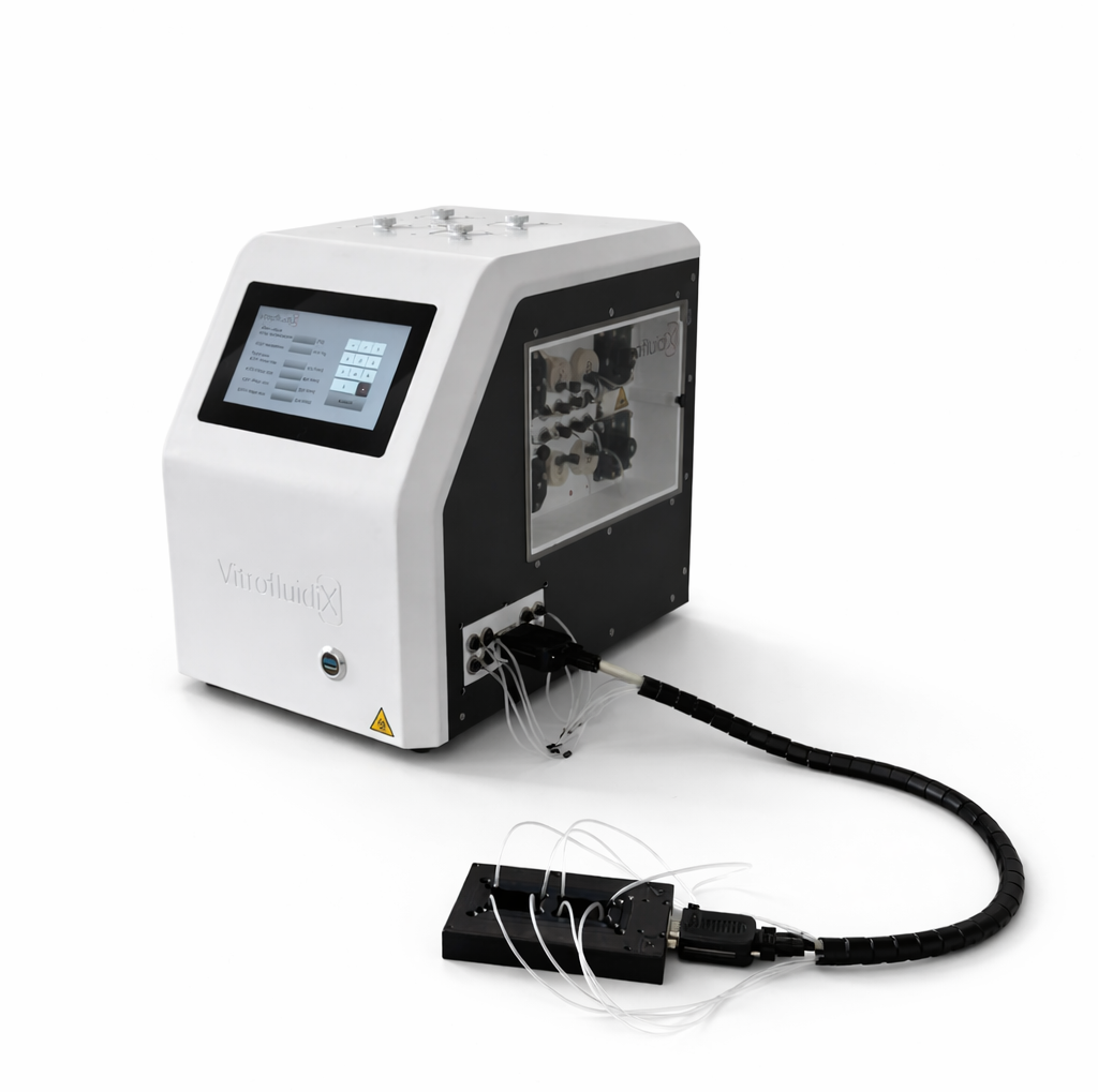
VitroFlow is not just a device – it is a fully integrated platform designed to enable scalable and reproducible organ-on-chip experiments.
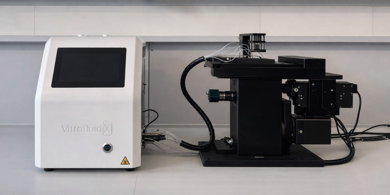
The platform combines controlled perfusion, incubation, live imaging and monitoring in a single system, while remaining compatible with a wide range of microfluidic chips and experimental setups.
Organ-on-chip experiments traditionally require multiple devices, fragmented software tools and complex laboratory setups.VitroFlow brings these components together into a single integrated platform.
VitroFlow is a unified, modular platform that integrates hardware, software, and consumables – while remaining fully compatible with existing lab infrastructure.
Plaftform Components
VitroFlow — Integrated perfusion, incubation and environmental control in a single system.
Free choice of chip and cells
Temperature control up to 60 °C (±0.1 °C accuracy)
24/7-Live-Monitoring
Customizable gas incubation at the microfludic scale
4 seperatly adjustable and analysable flows 2–1000 μL/min
Full Integration & Automation
Result for the customer
- Reproducible results across people and labs
- Less hands-on time
- Fewer errors and reliable routine operation in multi-user environments
Chip-Agnostic Interoperability
Result for the customer
- Maximum scientific freedom and collaboration compatibility
- Easier tech transfer between companies & universities
- Keep your current chips and models (no redesign to fit one vendor’s consumables)
Live Observation (Real-Time Readouts)
Result for the customer
- Access the full spectrum of microscopy (widefield, confocal, high-content, etc.)
- Enable advanced imaging-driven assays (e.g., time-lapse, 3D, multiple endpoints)
- Get more data per experiment by adding imaging modalities/readouts
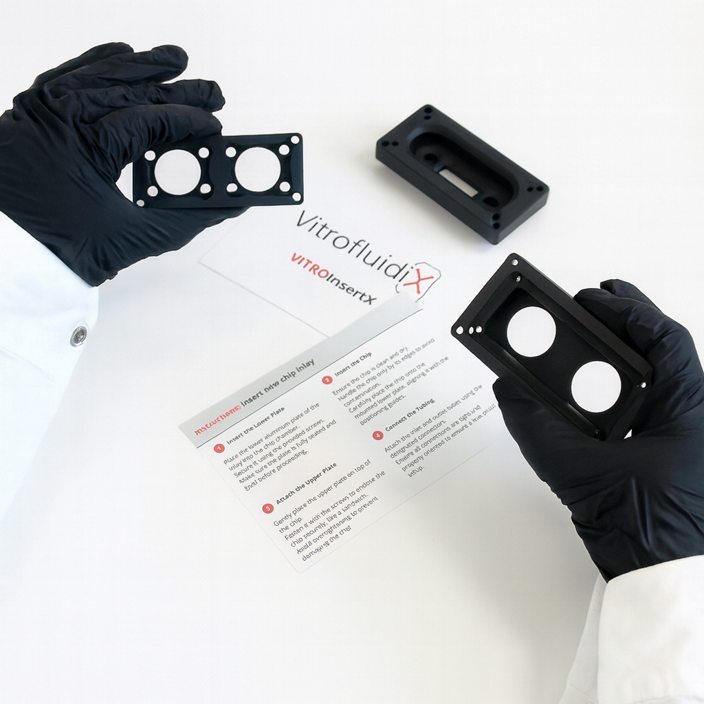
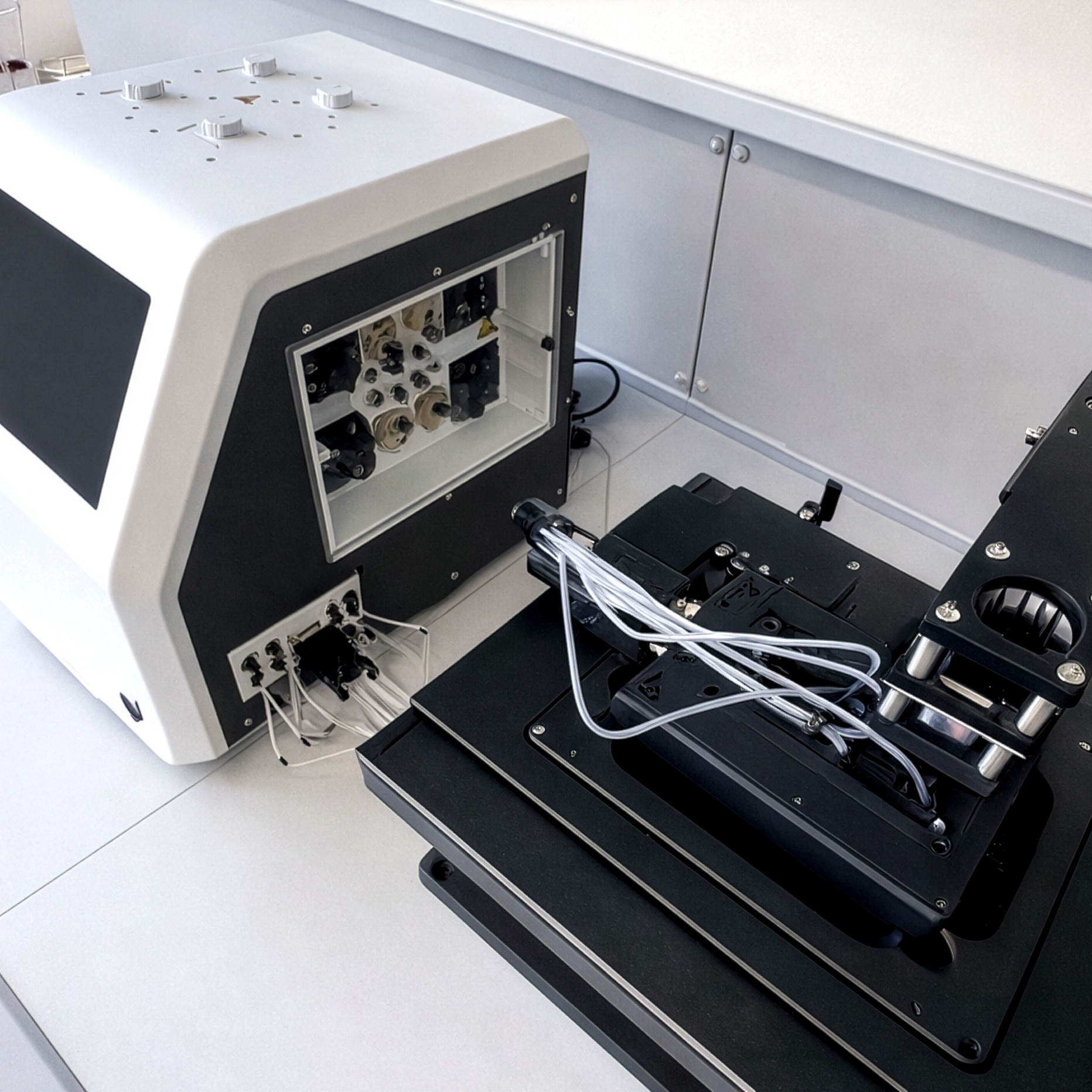
Fully Compatible and Customizable Chamber System
The chamber is fully mobile and compatible with standard microscope stages, enabling high-resolution imaging and screening. A microfluidic chip is clamped between two inlay parts. The inlays are fully customizable, allowing precise adaptation to a wide range of chip types – from commercially available formats to proprietary designs.
- ANSI/SLAS standard plate
- Dimensions for microscopy and screening
- Integrated heating for chip temperature control
- Customizable insert for different chip formats
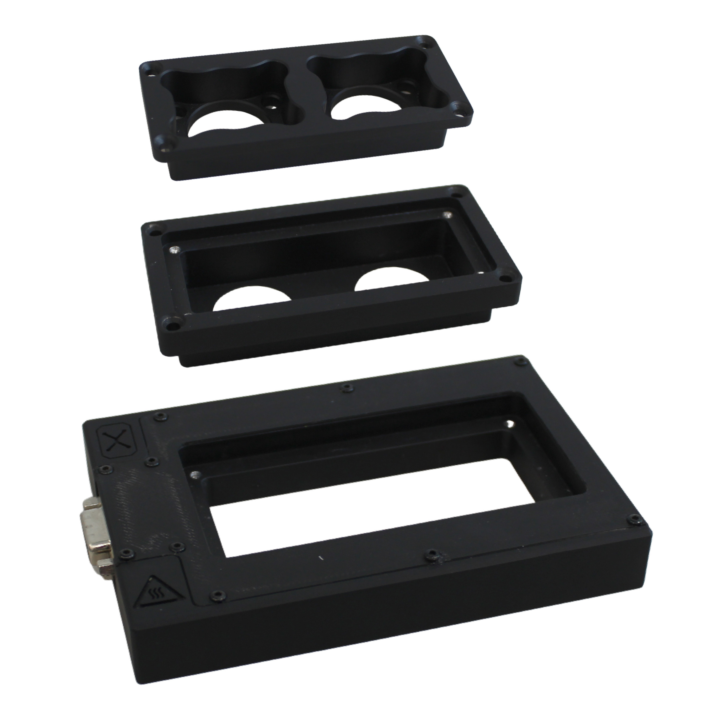
The upper inlay securely holds the chip in place and can be customized to match different chip geometries.
The lower inlay provides stable support for the chip and ensures precise alignment during imaging and screening.
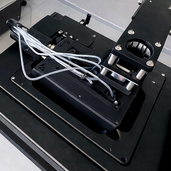
The base serves as the foundation of the chamber, housing integrated functions such as heating and compatibility with standard microscope stages.
Flexible chip formats for different experimental setups
Use the chip architecture that fits your research – no platform lock-in.
Supported formats
- Single microfluidic chips
- Multi-organ chip systems
- 2D and 3D cell models
- Barrier models
- Custom geometries
Experimental flexibility
- Compatible with different suppliers
- Integration of existing chip designs
- Flexible configurations
- Single or multiplexed experiments
Maximum control. Minimal Presence.
Our app connects directly to VitroFlow.Bio & allows you to monitor and control your experiments – automated, remote & smart.
VitroFlow.Connect supports the analysis and documentation of microfluidic cell culture experiments – intuitively, reliably and efficiently. Live imaging is automated and data storage is simplified.
- Less manual intervention – lower error rate
- Better collaboration across institutes
- Basis for future AI evaluation tools
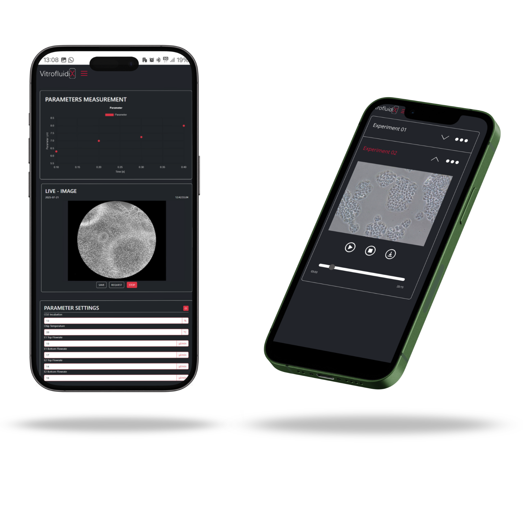
The app continuously records, displays, and saves live sensor data (flow rate, temperature & gas concentration)
App automatically captures and saves images throughout the experiment.
Live & automatic
We support in integrating your chips, prepare your experimental setup – while offering compatibility with standard microscopy systems!
All data, always at hand
Sensor values (temperature, flow rate, etc.) are displayed & saved.
Work & analyse remotely
Remote control of experiment parameters & access to saved protocols – ideal for weekends & cross-location teams.
More data. Better insights.
Compatible with standard microscopes and enables multi-level readouts – from live imaging to functional and kinetic analysis.
For the first time: More information per experiment
- No more “black box” experiments
- Learn real-time, not later
- Fewer experiments for the same answers
Live sensors, imaging today. Designed for modular extension.

Procedure of an experiment
with VitrofluidiX
Define Your Setup
First, you select or create the protocol: define which medium to use, set flow parameters, and assign organ-specific presets. Ready-made templates for common organ models are available, or you can customize your own. Then, simply fill the designated media compartments in the system.
Insert the Organ-on-a-Chip
Now it’s time to place your chip. Seed the required organ-specific cells onto the chip of your choice – single-organ, dual-organ, or a custom model. Once prepared, insert the chip into the incubation chamber and connect it to the system.
Start the Experiment
With all components in place, you’re ready to launch. Start the protocol and let VitroFlow take over – the organ simulation begins, fully automated and precisely controlled.
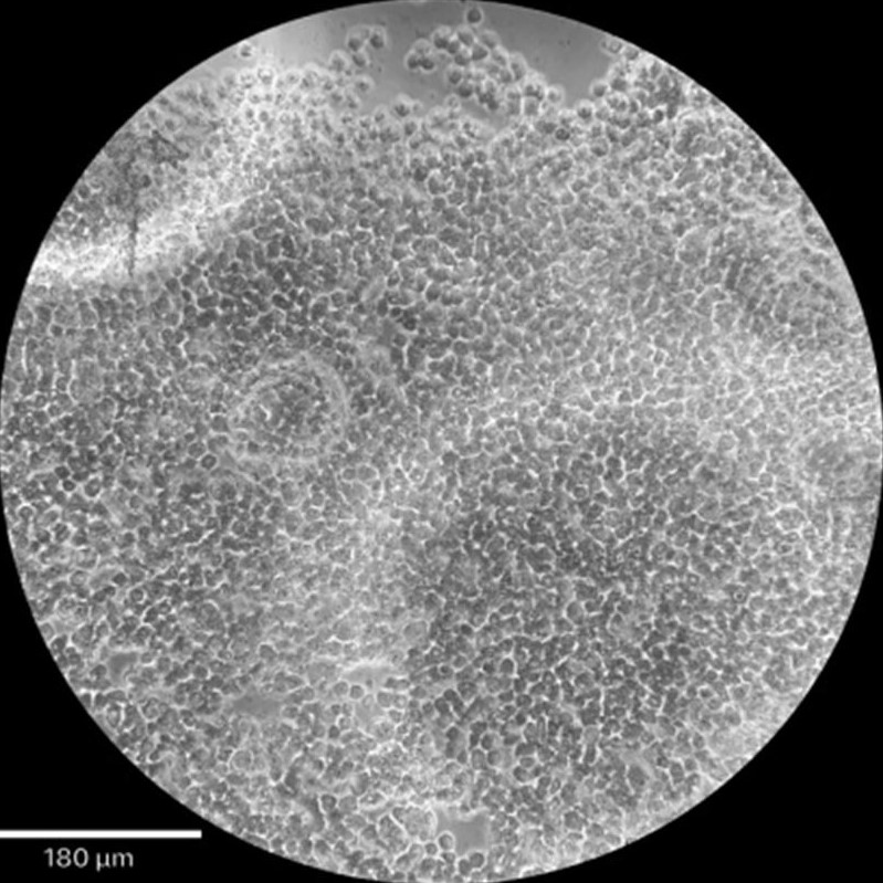
Analyze in Real Time
VitrofluidiX enables live imaging in fluorescence and phase contrast using microscopes already in your lab. The chip chamber follows the standard well plate format and can stay under the microscope throughout the experiment. Imaging runs continuously and automatically – ensuring real-time insights with no manual steps.
Designed to integrate seamlessly into existing lab workflows – reducing setup complexity and enabling faster experiment execution.