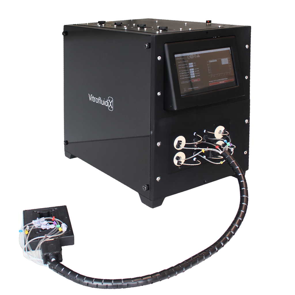
Organ-on-a-Chip made easy with VitroFlow.
- Cost-effective in vitro simulation of human organ function
- Live microscopy during the experiment
- Single & Multi-Organ Chips
- Cloud-based app for automated live imaging of cells and more
- Free choice of cells and chips
Why choose
VitroFlow?
VitroFlow creates a physiological microenvironment for human cell culture to study cellular interaction, morphological changes and responses toexternal stimuli under physiologically relevant biomechanical conditions for advanced drug screening and disease modelling.
Free choice of chip and cells
Temperature Control 25–90 °C
24/7-Live-Monitoring
Customizable gas incubation at the microfludic scale
4 seperatly adjustable and analysable flows 2–1000 μL/min
All in one OoC solution — all systems integrated for creating a physiologically relevant microenvironment
Full reproducibility — all parameters monitored and controlled by sensors.
24/7 live monitoring — use of any microscope (phase contrast & fluorescence) possible
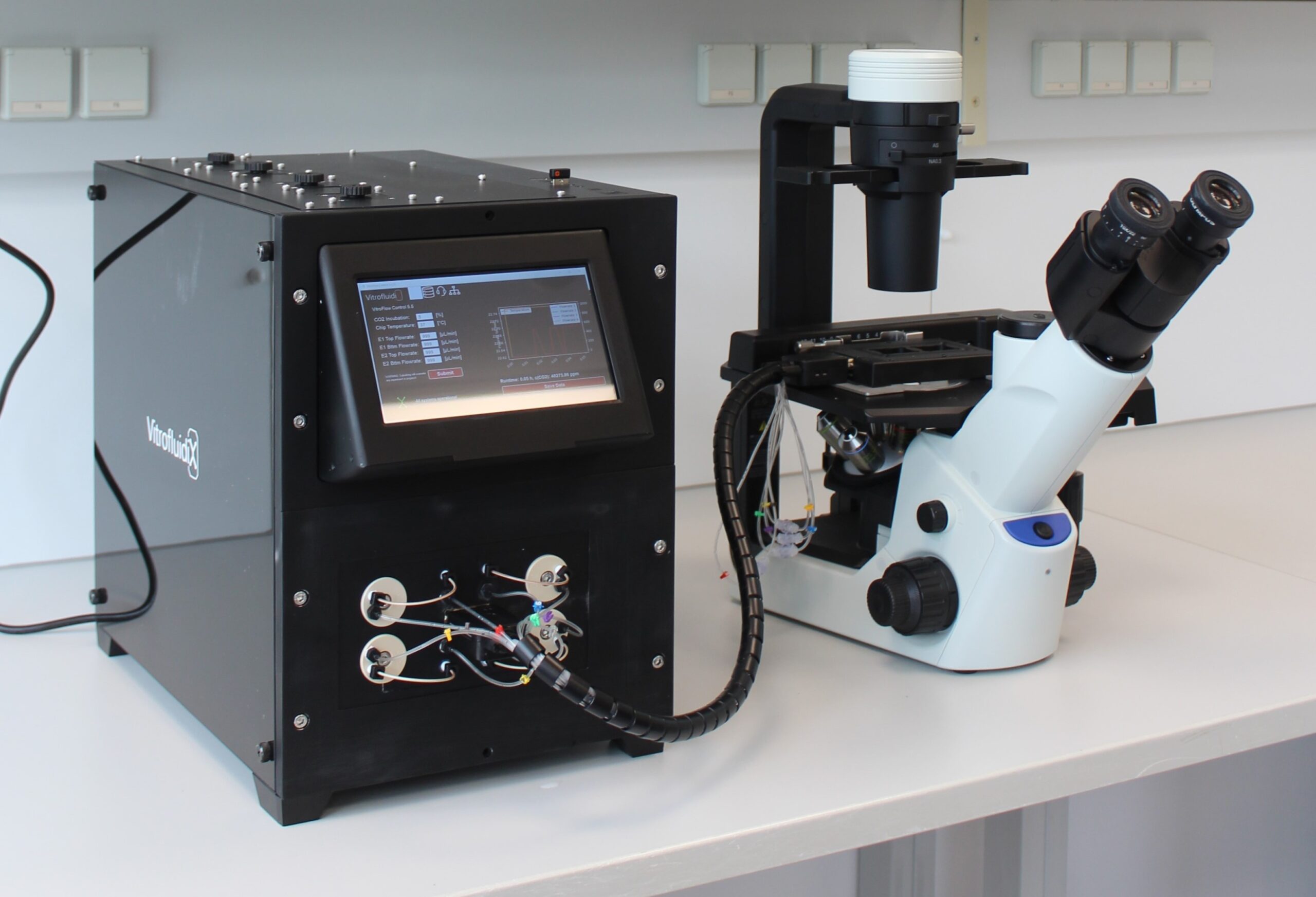
Fully Compatible and Customizable Chamber System
The chamber is fully mobile and compatible with standard microscope stages, enabling high-resolution imaging and screening. A microfluidic chip is clamped between two inlay parts. The inlays are fully customizable, allowing precise adaptation to a wide range of chip types—from commercially available formats to proprietary designs.
- ANSI/SLAS standard plate
- Dimensions for microscopy and screening
- Integrated heating for chip temperature control
- Customizable insert for different chip formats
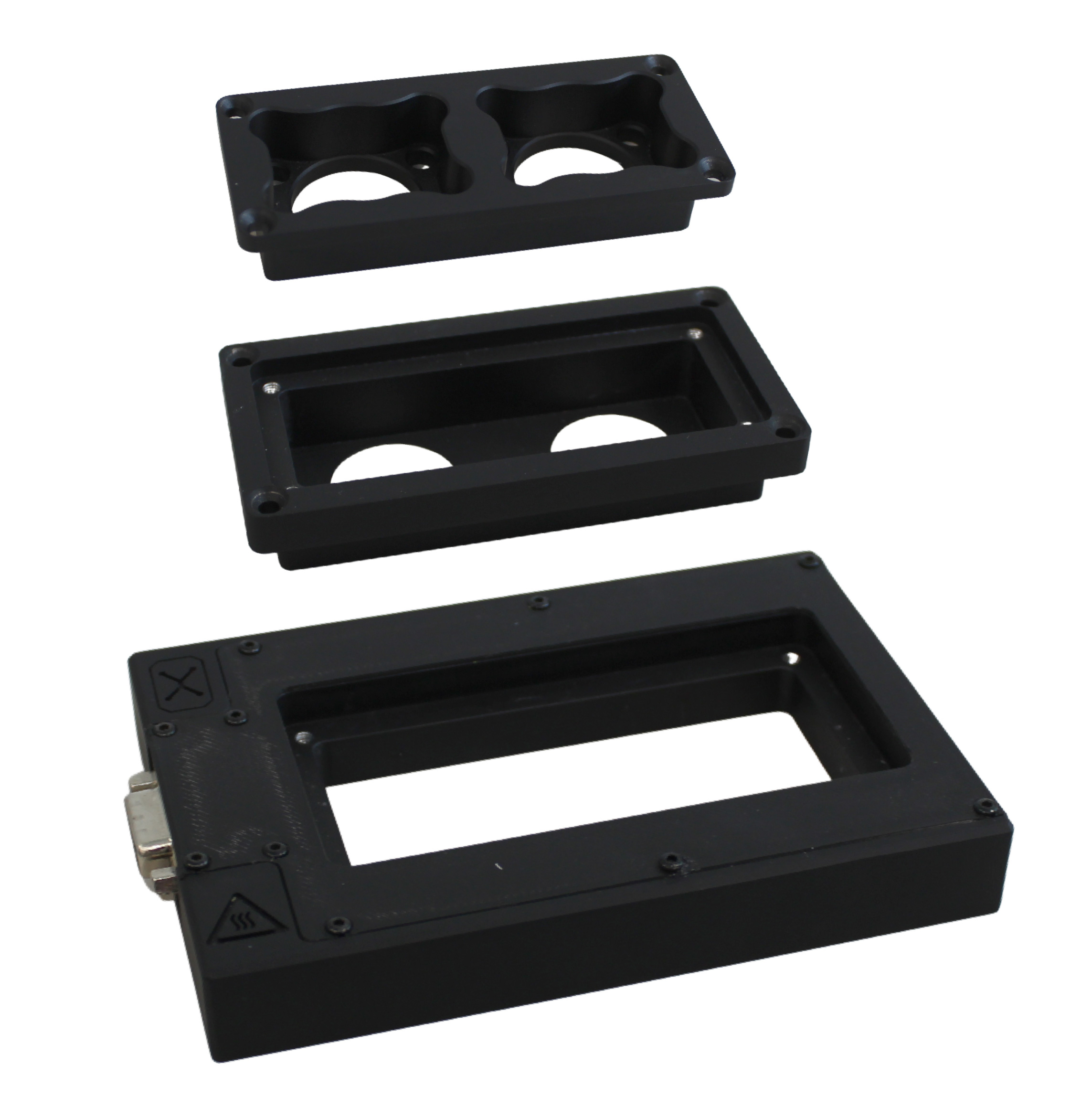
The upper inlay securely holds the chip in place and can be customized to match different chip geometries.
The lower inlay provides stable support for the chip and ensures precise alignment during imaging and screening.
The base serves as the foundation of the chamber, housing integrated functions such as heating and compatibility with standard microscope stages.
Maximum control. Minimal Presence.
Our app connects directly to VitroFlow.Bio & allows you to monitor and control your experiments – automated, remote & smart.
VitroFlow.Connect supports the analysis and documentation of microfluidic cell culture experiments – intuitively, reliably and efficiently. Live imaging is automated and data storage is simplified.
- Less manual intervention - lower error rate
- Better collaboration across institutes
- Basis for future AI evaluation tools
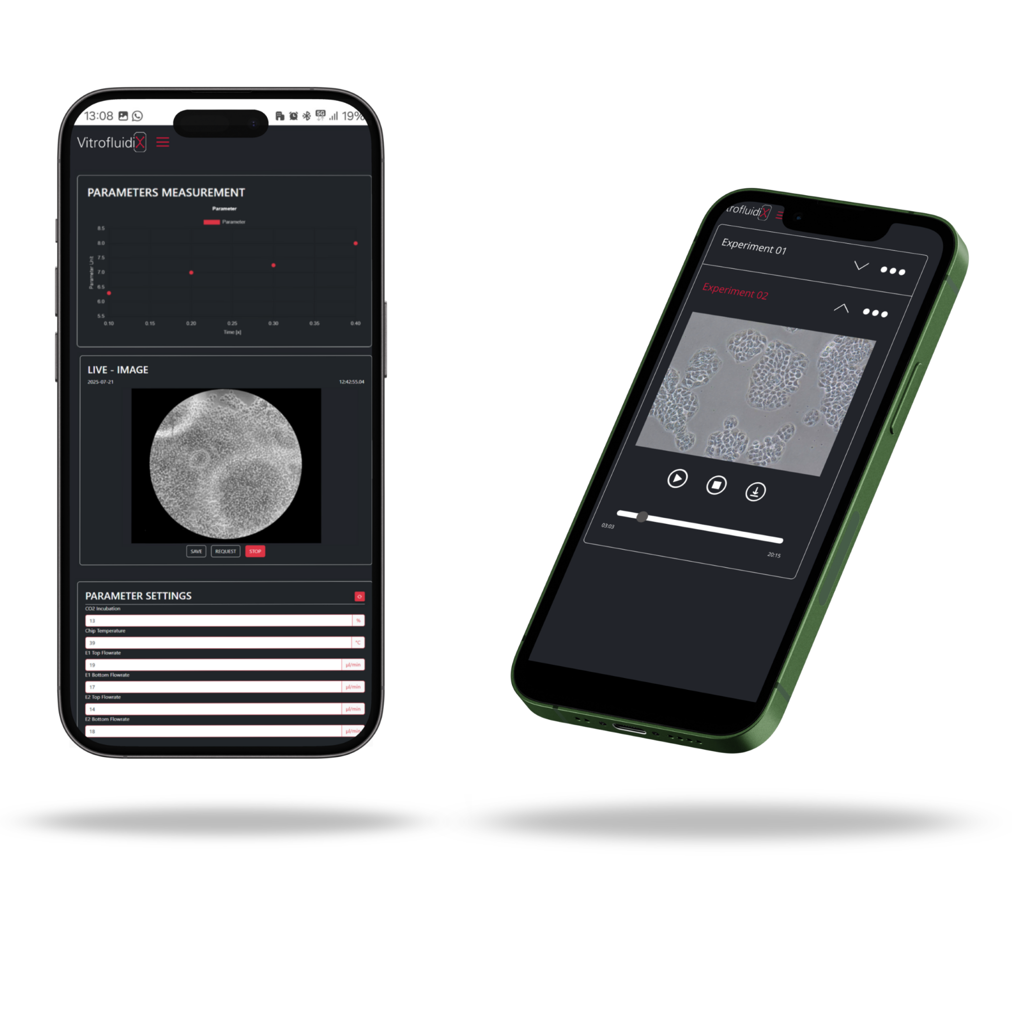
The app continuously records, displays, and saves live sensor data (flow rate, temperature & gas concentration)
App automatically captures and saves images throughout the experiment.
Live & automatic
Camera transmission in real time, storage of cell images at adjustable intervals
All data, always at hand
Sensor values (temperature, flow rate, etc.) are displayed & saved.
Work & analyse remotely
Remote control of experiment parameters & access to saved protocols – ideal for weekends & cross-location teams.
Procedure of an experiment
with VitrofluidiX
Define Your Setup
First, you select or create the protocol: define which medium to use, set flow parameters, and assign organ-specific presets. Ready-made templates for common organ models are available, or you can customize your own. Then, simply fill the designated media compartments in the system.
Insert the Organ-on-a-Chip
Now it’s time to place your chip. Seed the required organ-specific cells onto the chip of your choice — single-organ, dual-organ, or a custom model. Once prepared, insert the chip into the incubation chamber and connect it to the system.
Start the Experiment
With all components in place, you’re ready to launch. Start the protocol and let VitroFlow take over — the organ simulation begins, fully automated and precisely controlled.
Analyze in Real Time
VitrofluidiX enables live imaging in fluorescence and phase contrast using microscopes already in your lab. The chip chamber follows the standard well plate format and can stay under the microscope throughout the experiment. Imaging runs continuously and automatically—ensuring real-time insights with no manual steps.
Kits and accessories to power
your VitroFlow system

VitroInsertX
VitroInsertX is a modular inlay that fits chips from various manufacturers, enabling standardized handling and automation. It can be tailored to specific chip formats and offers full customization – from geometry to surface treatments.
VitroInsertX
VitroInsertX is a modular inlay that fits chips from various manufacturers, enabling standardized handling and automation. It can be tailored to specific chip formats and offers full customization – from geometry to surface treatments.

VITROKit RenewFlow P4
VITROKit RenewFlow P4 includes new pumping elements and spare connectors to maintain the flow unit of your VitrofluidiX Organ-on-Chip
VITROKit RenewFlow P4
VITROKit RenewFlow P4 includes new pumping elements and spare connectors to maintain the flow unit of your VitrofluidiX Organ-on-Chip

VITROKit Renew M5
VITROKit Renew M5 provides replacement membranes for the integrated bubble trap in the VitrofluidiX OOC system. Regular membrane exchange ensures reliable fluid handling and protects sensitive tissues from gas intrusion.
VITROKit Renew M5
VITROKit Renew M5 provides replacement membranes for the integrated bubble trap in the VitrofluidiX OOC system. Regular membrane exchange ensures reliable fluid handling and protects sensitive tissues from gas intrusion.

VITROKit 100
VITROKit 100 is optimized for ongoing operation of the VitrofluidiX OOC device. Recommended for 1xc use every 3 month, it supplies key consumables such as vials and crimp components to ensure seamless replenishment of cell culture media during routine workflows.
VITROKit 100
VITROKit 100 is optimized for ongoing operation of the VitrofluidiX OOC device. Recommended for 1xc use every 3 month, it supplies key consumables such as vials and crimp components to ensure seamless replenishment of cell culture media during routine workflows.

VITROKit 100+
VITROKit 100+ contains all components needed to start operating VitroFlow.Bio. It includes vials, crimp covers and plugs, and the appropriate crimping tool. Designed for a smooth system setup and reliable cell culture media handling from day one.
VITROKit 100+
VITROKit 100+ contains all components needed to start operating VitroFlow.Bio. It includes vials, crimp covers and plugs, and the appropriate crimping tool. Designed for a smooth system setup and reliable cell culture media handling from day one.